Good afternoon, Prof. Lo Chung Mau, Secretary for Health, Health Bureau, Prof. Gilberto Leung, President Hong Kong Academy of Medicine, Dr. the Honorable David Lam, Legislative Councillor, Legislative Council, Dr. Liza To, Assistant Director of Health, Department of Health, Dr. Law Chun Bon, Kowloon West Cluster Cluster Chief Executive, Hospital Authority, Prof. Peter Schirmacher, President of the European Society of Pathology, Dr. Raed Al Dieri, Chief Executive Officer of the European Society of Pathology Presidents and representatives from Sister Colleges, Fellows, Members, Associates, ladies and gentlemen.
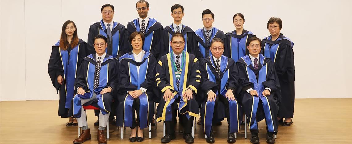
May I welcome you all to the conferment ceremony of our College this afternoon.
Congratulations:
Today is a very special day for our new Fellows. This marks a new milestone to recognize your tremendous effort and hard work over the past few years. Congratulations! I would also like to invite everyone in the audience to give your family, including your parents, significant others and your children, a round of applause for their unwavering support and encouragement. It is important to thank your mentors, trainers and colleagues for their guidance, as well as your patients, whom you have helped manage, for providing you with valuable training opportunities.
For those Members in the audience who have already passed their intermediate exam, I am sure it will soon be your turn for Fellowship conferment.
CME/CPD reminder and Genetic and Genomic Pathology:
To our new Fellows: Your future practice requires you to be competent in your field of practice. CME/CPD, continuing medical education/continuous professional development, is essential for your career development. As a usual practice of our College, I shall leave this part to Prof. Leung and I believe Prof. Leung from the Academy will remind you again later.
Hong Kong Academy of Medicine Faculty Development:
Following the Academy's Position Paper published in 2023, our College has committed to aligning with the Academy's latest Faculty Development initiatives. The Academy has organized several activities this year, including the 1st and 2nd Competency-based Medical Education Seminars, Basic Medical Education Courses, and Learner onLine Educator Courses. The purpose of these training events is to create a momentum for future faculty development in post-graduate medical education. A number of our College's key members have already joined these activities, and this would form the foundation for our College to follow the Academy's direction in Faculty Development.
There are several domains in the perspective of Faculty Development, including the structure of training and involvement of relevant stakeholders. Last year, we revised the Regulations to incorporate workplace-based assessments in various disciplines. Led by the Training and Examinations Committee, we are moving forward to engage College key members, Educational Supervisors, trainers and, most importantly, trainees in further enhancing training outcome. We believe that there are a number of skills our trainers need to acquire in order to ensure improved training outcomes. I hope that our trainees, who will eventually become one of our trainers in the College, will also actively engage in providing feedback on the training process.
Digital Pathology and Artificial Intelligence:
Talking about the Regulations, our College has recently revised and published the latest version of the training Regulations. The College has incorporated the concept of digital pathology in the latest regulations. I believe most of us have witnessed the rapid evolution and advancement in digital pathology over the past year and might have heard about various proposals in adoption of artificial intelligence in the field of Pathology. I would also like to report that the College Council has just endorsed the latest Guidelines in Implementation of Digital Pathology and Adoption of Artificial Intelligence. Details will be announced in due course.
I believe most of us might have been questioned or challenged about whether Artificial Intelligence will replace the role of pathologists. This was also a question I pondered about when ChatGPT first rolled out. Learning from the recent Academy Medical Education Conference, I strongly believe that the technology of artificial intelligence will not replace the role of pathologists, but pathologists using artificial intelligence will replace those who do not. The role of pathologists is unique in that we serve as a bridge between various advanced technologies in the field of laboratory medicine and clinical application. Taking reference from the field of Genetic and Genomic Pathology, we witness it being fully integrated into our clinical practice, for example, in tumour genetics, haematolymphoid malignancy, inborn errors of metabolism and immunopathology. The adoption of artificial intelligence would enhance our future working efficiency and the quality of diagnostic services. The adoption of artificial intelligence in image analysis and big data analysis may provide an edge in the next wave of technological advancement.
Green Pathology:
In addition, the implementation of the digital pathology aligns with another wave of recent development in the field of Pathology—Green Pathology. Having discussed with overseas colleagues across the globe, we realised that our College has various aspects to catch up on regarding environmental sustainability. The implementation of digital pathology represents one of the important steps to enhance our digital workflow in our daily practice. The College shall consider adopting a proactive approach to recent developments in the field of Pathology, with Green Pathology being one of the key directions.
European Society of Pathology:
One of our College's objectives is to promote advancement of knowledge in the science and art of pathology. In order to strengthen the role in promoting educational activities, our College signed a Memorandum of Understanding (MoU) with the European Society of Pathology earlier this year. I believe that this MoU would set the cornerstone for future international cooperation and foster further collaboration in the field of Pathology. I highly recommend that our College Fellows and trainees, particularly those in the specialties of Anatomical Pathology and Genetic and Genomic Pathology, learn about the various educational activities organised by the European Society of Pathology.
About dinner:
This year, our College is honoured to welcome prestigious guests from our European counterparts to join our Annual Dinner this evening. We encourage our College Fellows, Members and Associates to stay for the T.B. Teoh Foundation Lecture on "Third generation molecular pathology – from research to diagnostic implementation" by our Honorary Fellow Prof. Peter Schirmacher, followed by our Annual Dinner.
Hong Kong Academy of Medicine President Prof. Gilberto Leung:
I would also like to take this opportunity to express my sincere thanks to Prof. Gilberto Leung. Back in 2017, when I represented the College at the Academy's Young Fellows Chapter, I only marginally fulfilled the requirements to be a Young Fellow. Fortunately, it has been a privilege to have joined the Academy's Education Committee. Since then, I have had a number of opportunities to learn from Prof. Leung about the Academy's operation and participate in activities organised by the Academy. This invaluable experience has greatly benefited my career. Thank you, Prof. Leung.
Lastly, I would like to express my gratitude to all those who have contributed to the preparation of this event. It has been a lot of hard work, as always. I wish everyone the best in the days to come.

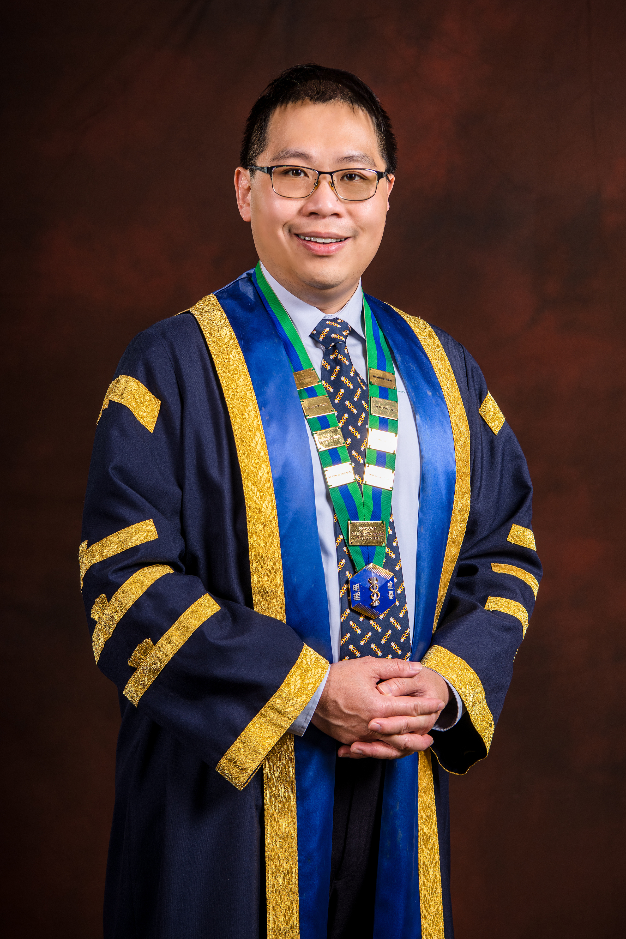
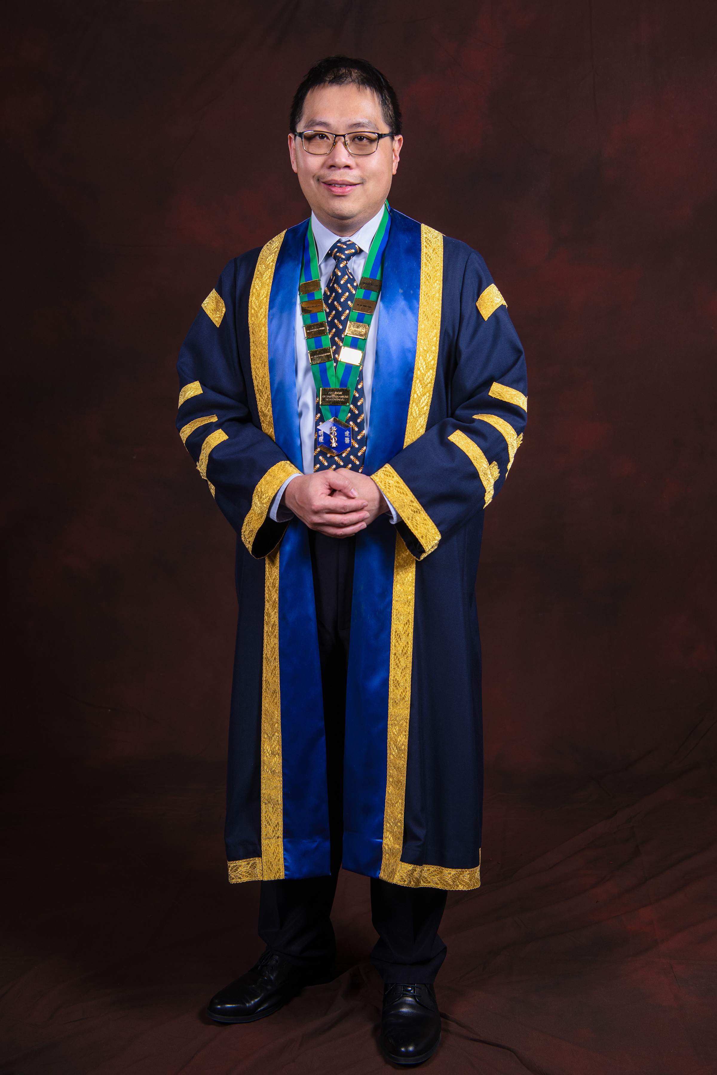
Dr MAK Siu Ming
President
16 November 2024